

From anywhere in the world
Tehran clinic services
An ovarian cyst containing hair and dental tissue can have a striking appearance. These findings have a recognized biological explanation: an ovarian dermoid cyst develops from the patient’s own germ cells, which can form different types of tissue.
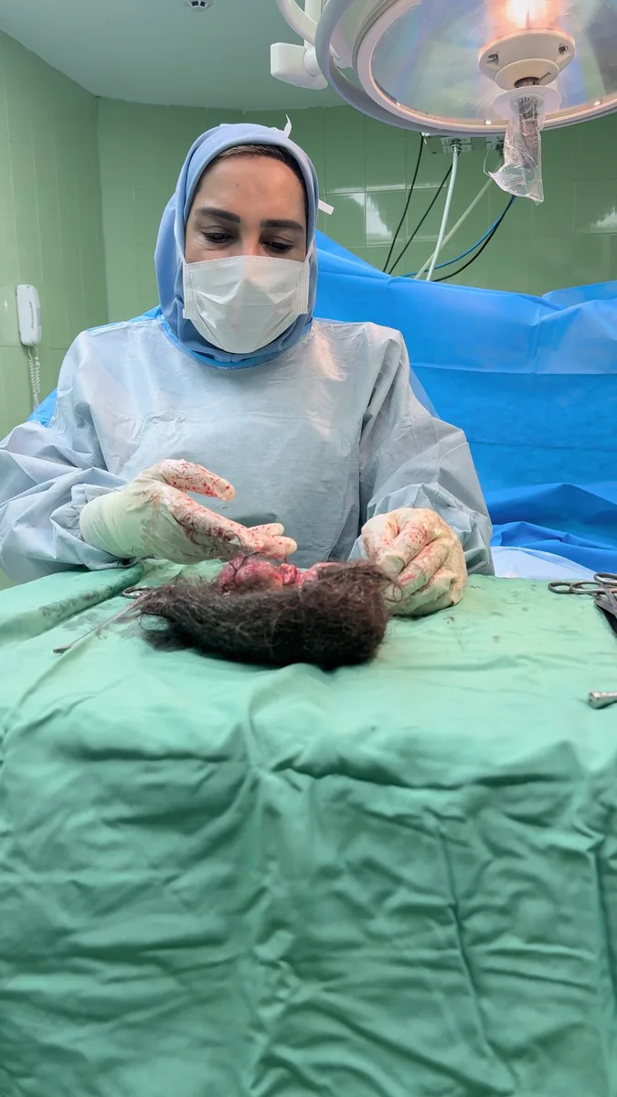
In a recent surgical case, Professor Mojgan Karimi-Zarchi, a full professor at Iran University of Medical Sciences and a gynecologic oncologist, removed a mass she identified as an ovarian dermoid cyst while preserving and repairing the ovary. The case provides an opportunity to explain both the unusual-looking contents and the clinical considerations involved in ovarian preservation.
The surgical findings
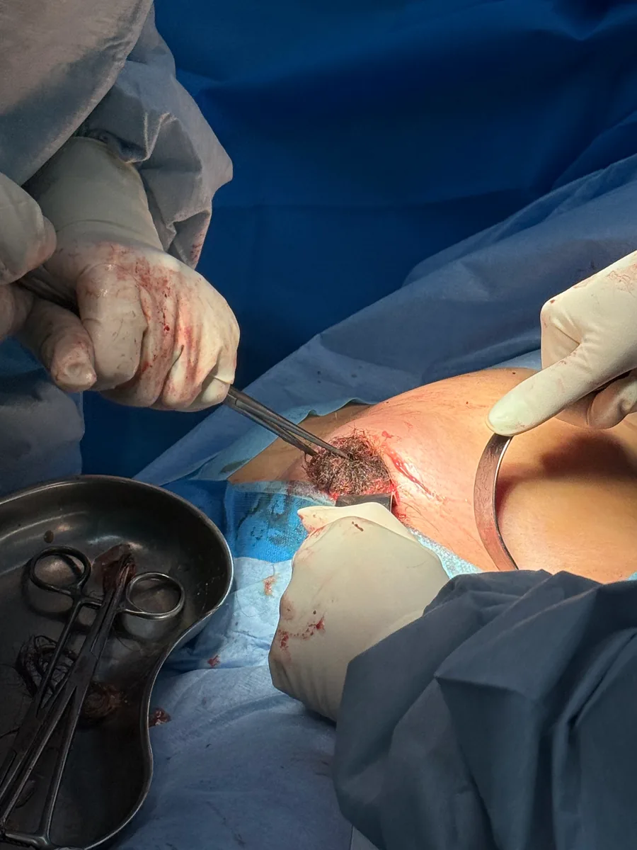
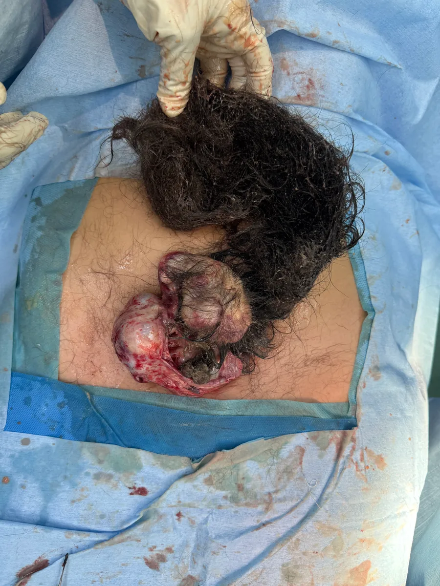
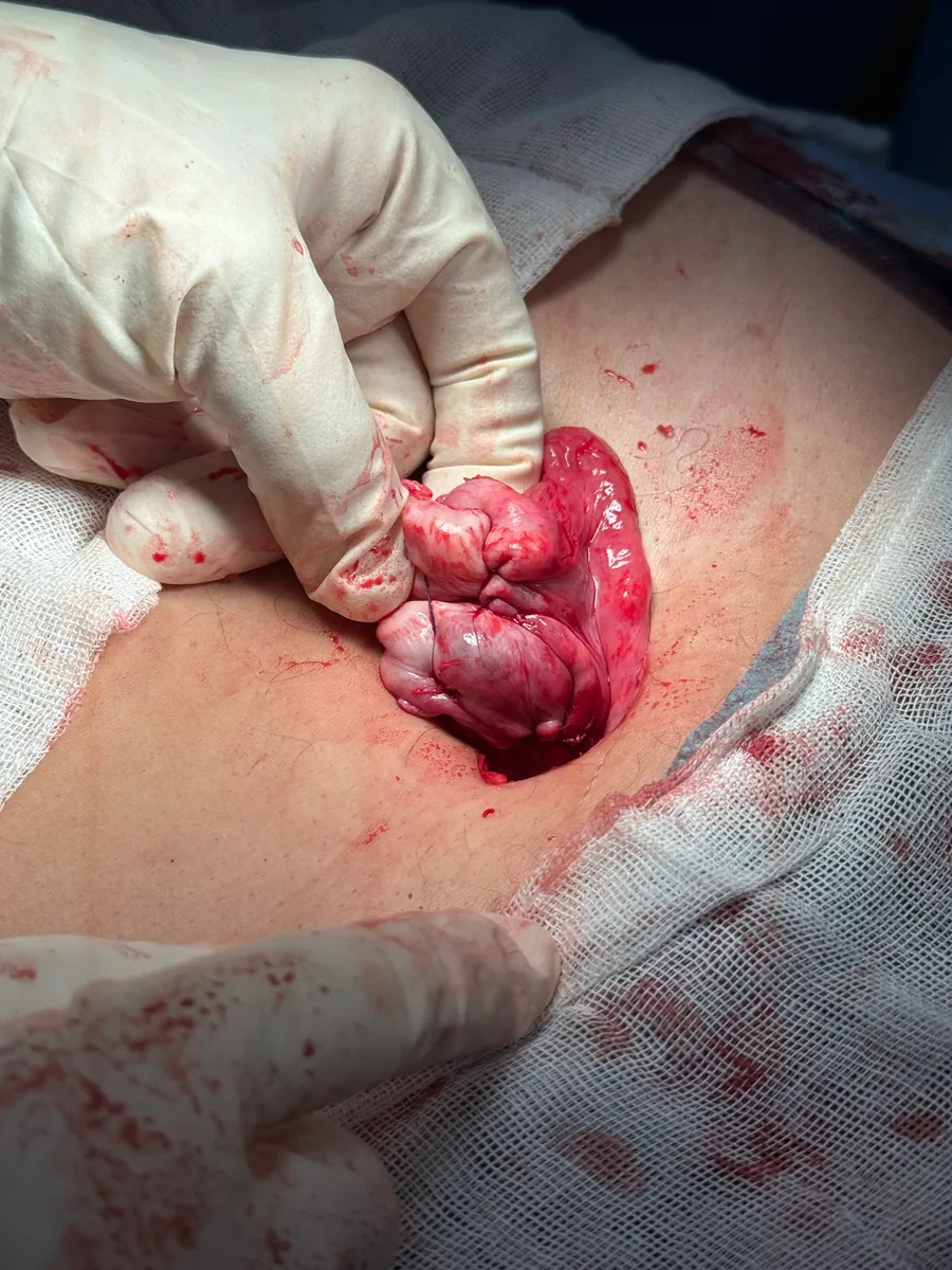
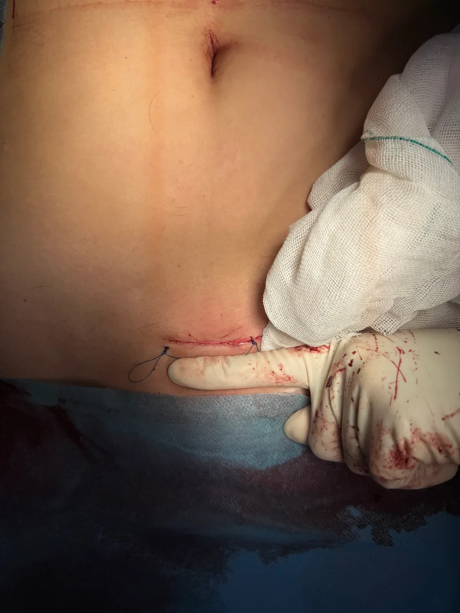
In her account of the operation, Professor Karimi-Zarchi identifies prominent hair and structures resembling teeth and bone within the removed mass. The photographs that follow show the ovary after repair and the surgical incision.
The central surgical finding is the removal of the mass with retention of the ovary. The appearance of the specimen explains the public interest in this case; the repaired ovary gives the operation its clinical context. This account concerns the operative findings and the surgeon’s explanation. It does not present a histopathological diagnosis or long-term ovarian-function results.
This section contains real surgical images.
Why can an ovarian cyst contain hair and teeth?
An ovarian dermoid cyst is also known as a mature cystic teratoma. It belongs to the group of ovarian germ-cell tumors. Germ cells are associated with the formation of eggs and have the capacity to develop into different tissue types. In a dermoid cyst, this can produce skin, hair, fatty material, teeth or bone.
The word “mature” describes the differentiation of the tissues within the tumor. It does not mean that the mass is a developing baby, and the presence of hair or teeth is not evidence of pregnancy. These contents arise from the patient’s own cells. They are recognized features of dermoid cysts, although their amount and appearance vary between cases.
How is a dermoid cyst assessed?
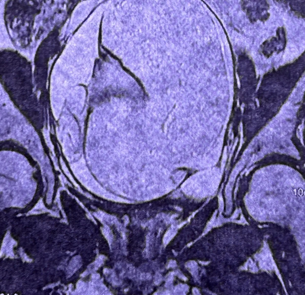
Assessment combines the patient’s symptoms and medical history with examination and imaging. Ultrasound is commonly used to evaluate an ovarian mass. Its appearance can suggest a dermoid cyst; MRI may help characterize a mass when further clarification is needed.
Most mature cystic teratomas are benign. The unusual appearance of hair or teeth does not, by itself, indicate cancer. Equally, a photograph cannot establish the final tissue diagnosis. When a mass is removed, histopathological examination establishes the tissue diagnosis and assesses for malignancy.
A dermoid cyst also differs from a functional cyst related to the menstrual cycle. The expectation that some functional cysts disappear over a few cycles should not be applied to a dermoid cyst.
Removing the cyst while preserving the ovary
A cystectomy removes the cyst while retaining ovarian tissue. An oophorectomy removes the ovary. The choice depends on the clinical assessment, the characteristics of the mass, the amount of ovarian tissue that can be retained, and the patient’s circumstances and reproductive plans.
When appropriate, preserving ovarian tissue is an important consideration. It must be balanced with safe removal and the findings at surgery. The approach used for one patient cannot automatically be applied to every ovarian mass.
In the case described here, Professor Karimi-Zarchi reports that the ovary was retained and repaired after removal of the mass. This documents anatomical preservation during surgery. Ovarian function and future fertility are separate outcomes that require clinical assessment and follow-up; they cannot be determined from the postoperative appearance alone.
Does every dermoid cyst need surgery?
Management is individual. Selected cysts can be monitored, while symptoms, growth, concern about complications or uncertainty about the nature of the mass may favor surgery. Observation means a planned follow-up strategy with a clinician, rather than assuming the cyst will disappear.
Dermoid cysts may be found incidentally or cause pelvic discomfort. Complications can include twisting of the ovary, called torsion, or rupture. These possibilities help explain why a known cyst needs an appropriate clinical plan, even when it is thought to be benign.
Can it be prevented, and when should symptoms be assessed?
There is no established method of preventing an ovarian dermoid cyst from forming. A screening scan does not prevent one. Ultrasound may be useful for assessing symptoms or following a known cyst; reading about a surgical case does not, on its own, establish a need for imaging.
Persistent pelvic pain, ongoing bloating, increasing abdominal swelling or feeling full after eating small amounts should be discussed with a clinician. These symptoms can have several causes and are not specific to a dermoid cyst.
Sudden, severe pelvic pain, or abdominal pain accompanied by nausea or vomiting, requires urgent medical assessment. A person with these symptoms should not wait for a routine appointment.
About Professor Mojgan Karimi-Zarchi
Professor Karimi-Zarchi is a full professor at Iran University of Medical Sciences and a gynecologic oncologist. Her work spans more than two decades of clinical practice and teaching in women’s health, with a focus on gynecologic cancers, precancerous conditions and fertility-preserving care where appropriate.
This surgical case is shared as part of that clinical and educational work: to explain a recognized ovarian condition and the decisions involved in its treatment.
This article is for general education and does not replace an appointment with your physician. Reviewed: September 2026.